


Orders placed before 12 noon
are usually dispatched on the same day (no dispatch on Fridays)
Gram Staining Kit for Tissue Sections – Histology
EAN: 08057724950823
Article number
KGR.30
Packaging unit
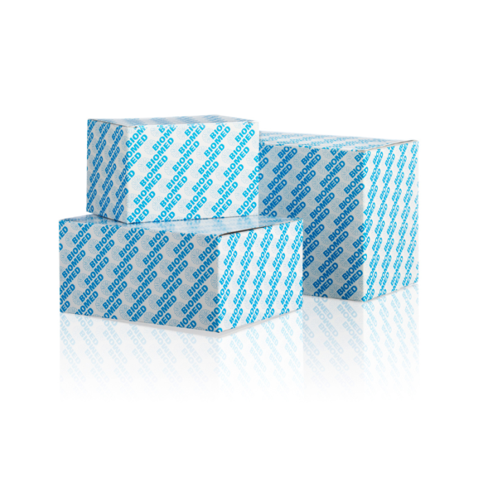
1 x 30 mL Reagent 1 Kristallviolett, 1 x 30 mL Reagent 2 Kaliumiodid/Iod, 1 x 100 mL Reagent 3 Aceton/Ethanol/Isopropanol, 1 x 30 mL Reagent 4 Ethanol/Isopropanol/basisches Fuchsin, 1 x 100 mL Reagent 5 Aceton/Ethanol/Isopropanol
Ready-to-use Gram staining kit for the light microscopic differentiation of Gram-positive and Gram-negative bacteria in histological tissue sections.
€93.80
Excl. 19% VAT ,
excl. Shipping Cost
Gram Staining Kit for Tissue Sections – Histological Bacterial Staining
The Gram Staining Kit for Tissue Sections provides ready-to-use staining reagents for the qualitative differentiation of Gram-positive and Gram-negative bacteria in histological tissue specimens. Designed for light microscopy, the kit enables bacterial structures to be visualized within their surrounding tissue architecture.
In histopathology, demonstrating bacteria directly in tissue sections can provide valuable additional information when investigating suspected bacterial infections. Unlike conventional microbiological smears, histological preparations allow microorganisms to be examined in relation to the surrounding cells and tissue structures.
The kit contains five reagent components for primary staining, iodine treatment, differentiation and counterstaining. The solutions are supplied ready to use and can be incorporated into validated manual staining procedures in medical laboratories.
Gram Staining of Tissue Sections in Histopathology
Gram staining is a well-established differential staining technique used to distinguish bacteria according to their staining characteristics.
In histological specimens, the method requires careful differentiation between bacterial structures and the surrounding tissue. Appropriate staining contrast is essential for microscopic assessment, particularly when microorganisms are present in small numbers or within complex tissue structures.
The Gram Staining Kit for Tissue Sections is intended for the qualitative in vitro differentiation of Gram-positive and Gram-negative bacteria in tissue sections of human and animal origin.
Tissue samples are fixed in formalin and embedded in paraffin. Before staining, sections are deparaffinized and rehydrated according to the laboratory's validated procedures.
Why Is Gram Staining Useful in Tissue Examination?
Histological Gram staining can support the microscopic assessment of bacterial structures in tissue specimens.
The technique allows stained microorganisms to be examined in their anatomical context, providing information about their distribution within the tissue.
Gram staining distinguishes bacteria according to their differential retention of the primary stain. However, it does not provide definitive identification at the species level.
A negative staining result does not exclude bacterial infection. The sensitivity of microscopic detection depends on factors such as bacterial abundance, specimen preparation and staining quality.
How Does Gram Staining Work in Histological Sections?
The Gram staining technique is based on the different abilities of Gram-positive and Gram-negative bacteria to retain the crystal violet–iodine complex during differentiation.
Initially, bacterial structures are stained with crystal violet. Subsequent treatment with an iodine solution forms a crystal violet–iodine complex.
During differentiation with an acetone–alcohol solution, the complex is removed from Gram-negative bacteria, whereas Gram-positive bacteria retain their blue-violet coloration.
Counterstaining with basic fuchsin subsequently produces an intense pink to fuchsia coloration in Gram-negative bacteria.
The resulting staining pattern enables the two bacterial groups to be distinguished by light microscopy.
Expected Staining Results
Gram-positive bacteria: Blue to violet
Gram-negative bacteria: Intense pink to fuchsia
Cell nuclei: Pink
Cytoplasm and background: Pale pink
Staining results should always be interpreted in conjunction with tissue morphology and the relevant clinical findings.
Five Ready-to-Use Reagent Components
The Gram Staining Kit for Tissue Sections contains five reagent components corresponding to the individual stages of the staining procedure.
Crystal Violet Solution – Primary Staining
Crystal violet provides the initial staining of bacterial structures and forms the basis of the Gram differentiation process.
Iodine-Iodine Solution – Dye Complex Formation
The iodine solution contains iodine and potassium iodide. It promotes the formation of the crystal violet–iodine complex within stained microorganisms.
Acetone-Alcohol Solution – Differentiation
The acetone–alcohol solution removes the primary dye complex from Gram-negative bacteria during the differentiation stage.
Correct differentiation is important for distinguishing Gram-positive and Gram-negative organisms.
Alkaline Fuchsin Solution – Counterstaining
Basic fuchsin provides the contrasting pink to fuchsia coloration of Gram-negative bacteria, making them distinguishable from Gram-positive organisms.
Acetone-Alcohol Solution – Final Differentiation
A further acetone–alcohol treatment is performed during the later stages of the staining procedure.
This step requires particular attention because the extent of differentiation may vary depending on the specimen.
All staining reagents are supplied ready to use, eliminating the need for reagent preparation before staining.
Manual Gram Staining of Histological Tissue Sections
The Gram Staining Kit for Tissue Sections is intended for use by appropriately trained laboratory professionals.
Before staining, formalin-fixed, paraffin-embedded tissue sections must be deparaffinized and rehydrated.
The manual staining procedure includes primary staining with crystal violet, iodine treatment, differentiation with acetone–alcohol and counterstaining with basic fuchsin.
In the described procedure, the crystal violet, iodine and fuchsin staining steps each have an incubation time of three minutes.
The differentiation stages and rinsing steps are performed in the specified sequence. Particular care is required during acetone–alcohol differentiation, as insufficient or excessive differentiation may affect the staining result.
After the final staining steps, sections are dehydrated, cleared and mounted for microscopic examination.
The staining procedure must be performed according to the laboratory's validated method.
Ready-to-Use Staining Solutions for Laboratory Workflows
Ready-to-use reagents simplify the preparation of histological Gram staining procedures.
By eliminating the need to prepare individual staining solutions, the kit can help laboratories organize routine staining workflows more efficiently.
Consistent staining results nevertheless depend on appropriate specimen preparation, correct reagent handling and careful control of the differentiation stages.
In particular, the differentiation process influences the contrast between Gram-positive and Gram-negative bacteria.
Regular inspection of staining solutions and adherence to established laboratory procedures are important for maintaining staining quality.
Gram Staining of Tissue Sections Versus Microbiological Smears
Gram staining is used in both histopathology and microbiology, but the specimen types and analytical objectives differ.
In histological tissue sections, the main objective is to visualize bacterial structures within the tissue architecture. The surrounding cells, extracellular structures and background staining must be considered during microscopic interpretation.
In conventional microbiological Gram staining, bacterial smears are commonly examined to assess bacterial morphology, arrangement and Gram reaction.
For microbiological staining applications, BIOMED also offers products within its Gram staining range.
Further staining reagents and laboratory staining methods are available in the BIOMED staining solutions range.
Storage Conditions and Shelf Life
The staining solutions must be stored protected from light at +15 °C to +25 °C.
Unopened bottles remain stable until the stated expiry date when stored under the specified conditions. Do not freeze.
After opening, tightly closed bottles may be used for up to two years when stored correctly, but never beyond the labelled expiry date.
The solutions must be inspected regularly for contamination.
Because the kit contains volatile substances, it must be used with a suitable extraction or ventilation system.
Gram Staining Kit for Tissue Sections – Product Specifications
Product: Gram Staining Kit for Tissue Sections
Application: Qualitative Gram staining of histological tissue sections
Specimen type: Tissue sections of human and animal origin
Method: Light microscopic differentiation of Gram-positive and Gram-negative bacteria
Reagent format: Ready-to-use staining solutions
Reagent components: Crystal violet, iodine-iodine solution, acetone–alcohol and alkaline fuchsin
Kit contents: 1 × 30 mL each of Reagents 1, 2 and 4; 1 × 100 mL each of Reagents 3 and 5
Storage: +15 °C to +25 °C, protected from light
Intended users: Trained medical laboratory professionals
Histological Gram Staining with Ready-to-Use Reagents
The Gram Staining Kit for Tissue Sections provides a complete set of ready-to-use reagents for the qualitative differentiation of Gram-positive and Gram-negative bacteria in histological preparations.
The staining procedure enables bacterial structures to be visualized by light microscopy and assessed in relation to the surrounding tissue morphology.
With dedicated reagents for primary staining, iodine treatment, differentiation and counterstaining, the kit can be integrated into validated histological staining workflows.
It is a practical choice for medical laboratories performing Gram staining of tissue sections as part of their histopathological examination procedures.
Frequently Asked Questions About Gram Staining of Tissue Sections
What is the Gram Staining Kit for Tissue Sections used for?
The kit is intended for the qualitative differentiation of Gram-positive and Gram-negative bacteria in histological tissue sections of human and animal origin using light microscopy.
Can Gram staining detect bacteria in tissue sections?
Gram staining can make stainable bacterial structures visible within tissue specimens. Detection depends on bacterial abundance, specimen preparation and the quality of the staining procedure.
What colors do Gram-positive and Gram-negative bacteria show?
Gram-positive bacteria appear blue to violet, while Gram-negative bacteria appear intense pink to fuchsia after counterstaining.
Are the staining reagents ready to use?
Yes. All staining solutions are supplied ready to use and do not require preparation before staining.
Which tissue specimens are suitable for this staining procedure?
The kit is intended for tissue sections of human and animal origin. Samples are formalin-fixed and paraffin-embedded, then deparaffinized and rehydrated before staining.
Can Gram staining identify the bacterial species?
No. Gram staining provides differential morphological information but does not allow definitive bacterial species identification.
How long can the staining solutions be used after opening?
When stored protected from light at +15 °C to +25 °C in tightly closed bottles, the solutions may be used for up to two years after opening, provided the labelled expiry date has not been exceeded. Regular inspection for contamination is required.
| Packaging unit | 1 x 30 mL Reagent 1 Kristallviolett, 1 x 30 mL Reagent 2 Kaliumiodid/Iod, 1 x 100 mL Reagent 3 Aceton/Ethanol/Isopropanol, 1 x 30 mL Reagent 4 Ethanol/Isopropanol/basisches Fuchsin, 1 x 100 mL Reagent 5 Aceton/Ethanol/Isopropanol |
|---|
Related Products
Check items to add to the cart or